
Mechanisms involved
This condition is explained through the mechanisms below. Each one is taught as ordinary physiology first.
Primary
Secondary / contributing
Inside the uterine wall
Adenomyosis — lining tissue trapped in the muscle
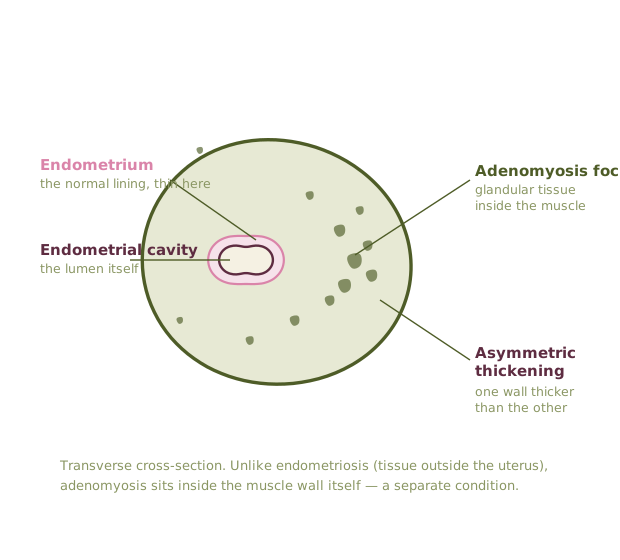
- Endometrium — the lining that sheds each cycle. Normally it stays inside the cavity.
- Adenomyotic foci — islands of lining-type tissue embedded in the muscle. They bleed and inflame with each cycle, with nowhere to drain.
- Myometrium thickens asymmetrically — the classic globular, tender uterus with heavy, crampy periods and pressure.
- Diagnosed on imaging (MRI or expert transvaginal ultrasound), not by laparoscopy — the opposite of endometriosis.
Schematic, not to scale. Adenomyosis lives inside the myometrium (the muscular wall), so the uterus becomes globular, boggy and tender, and periods get heavy. This is a different process from endometriosis, which grows outside the uterus.
Immune cells involvedWho is actually in the tissue
Cyclical bleeding inside muscle asks the immune system to clean up somewhere it cannot drain.
Selected cell
Macrophage
Clears debris and old blood. When clearance is incomplete it switches to a repair-and-inflame phenotype that feeds fibrosis and new nerve growth.
Iron-loaded macrophages accumulate around foci in the myometrium, sustaining low-grade inflammation and tenderness.
Schematic drawings, not micrographs. Immune findings here describe mechanism and association; none of them are diagnostic tests.
What it is
Glandular tissue is found within the myometrium itself, producing a bulky, tender uterus, heavy bleeding and diffuse deep pain. It is visible on high-quality transvaginal ultrasound or MRI, and it is a separate condition from endometriosis even though the two frequently coexist.
How the mechanisms produce this
- Cyclical bleeding within muscle produces edema, tenderness and heavy periods rather than the focal lesion pain of peritoneal disease.
- Junctional-zone disruption alters uterine contractility, which is felt as dragging, cramping pain distinct from lesion pain.
- Longstanding heavy bleeding drives iron deficiency, which independently worsens fatigue and orthostatic tolerance.
What tends to help
- Bleeding control first — it usually does more for daily function than anything else.
- Iron status checked and treated, ferritin as well as hemoglobin.
- Hormonal suppression, tranexamic acid, or an intrauterine device depending on goals.
- Distinguish which pain is uterine and which is peritoneal before deciding on surgery.
Where the evidence is thin
Prevalence is poorly established because diagnosis historically required hysterectomy, and imaging criteria are still being standardized.