
Mechanisms involved
This condition is explained through the mechanisms below. Each one is taught as ordinary physiology first.
Secondary / contributing
Outside the uterus
Endometriosis — implants on the peritoneum and organs
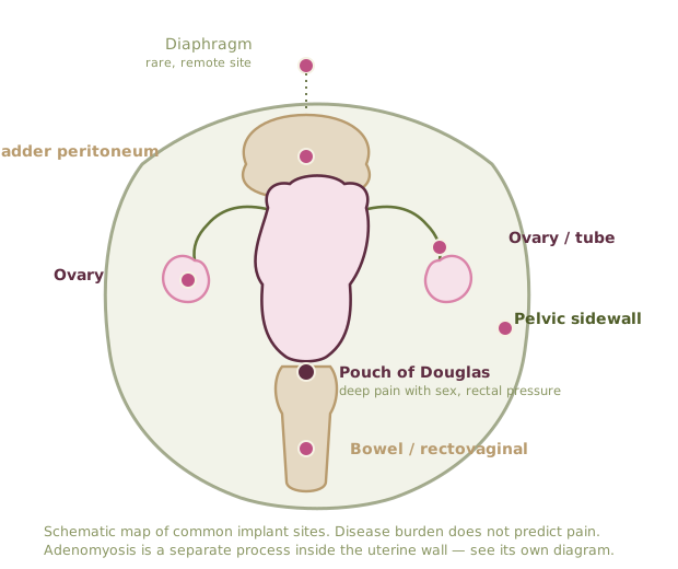
Schematic map of common implant sites. Disease burden does not predict pain. Adenomyosis is shown separately because it is a different process inside the uterine wall.
Immune cells involvedWho is actually in the tissue
Lesion tissue is not just misplaced — it sits inside an immune environment that lets it stay and keeps it painful.
Selected cell
Mast cell
Tissue sentinel packed with histamine, tryptase and prostaglandin granules. When it degranulates it recruits and sensitizes nearby nerve endings.
Lesions are dense in activated mast cells, sitting right against small nerve fibers. This is a large part of why lesion size and pain severity match so poorly.
Schematic drawings, not micrographs. Immune findings here describe mechanism and association; none of them are diagnostic tests.
What it is
Tissue resembling endometrium grows outside the uterine cavity — on peritoneum, ovaries, bowel, bladder, and occasionally further afield. It responds to hormonal signalling, provokes local inflammation and adhesions, and is diagnosed definitively at surgery, though imaging identifies deep and ovarian disease well.
How the mechanisms produce this
- Lesions carry estrogen and progesterone receptors and behave with the cycle, which is why pain often begins as cyclical and only later becomes constant.
- Lesion tissue is dense in mast cells and inflammatory mediators, which recruit and sensitize nearby small nerve fibers.
- Sustained nociceptive input from the pelvis is a textbook driver of central sensitization, which is why disease burden and pain severity correlate so poorly.
- Pelvic inflammation and pain both alter autonomic tone, contributing to the gut, bladder and orthostatic symptoms that ride alongside.
What tends to help
- Treat the lesion and the pain system in parallel — excision or suppression alone under-treats sensitized pain.
- Prostaglandin suppression started before pain onset in the cycle.
- Pelvic-floor physiotherapy where there is muscular guarding, which is most people by the time they are diagnosed.
- Explicit reassessment at three to six months rather than open-ended trials.
Where the evidence is thin
Recurrence rates after surgery vary widely by surgeon and by disease type, and no biomarker is ready for clinical use.